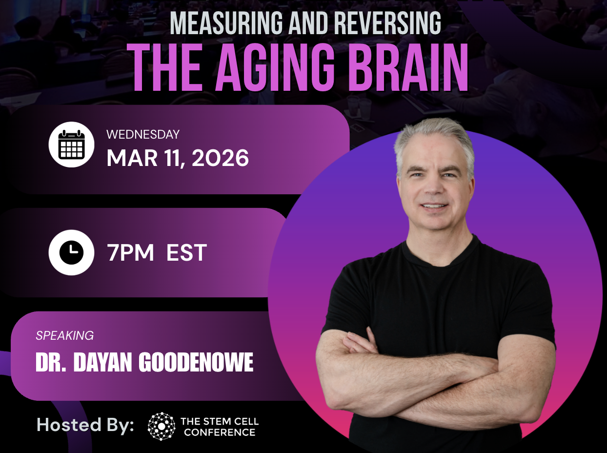
There is a graph of brain volume against age that Dr. Dayan Goodenowe put up early in our session, and the thing he wanted the room to notice was not the downward slope. It was that the top of the chart is empty. On his chart, nobody in their seventies or eighties had held onto full brain volume. Not one point.
Goodenowe, is a PhD rather than a physician, trained as a synthetic organic chemist and working on the biochemical mechanisms of neurological disease. He invented the non-targeted metabolomics technology behind his measurement work, wrote Breaking Alzheimer’s, and disclosed on the call that he founded the company producing the precursor compounds he studies. His argument is that brain aging can be measured with far more precision than most programs attempt, and that the measurements move.
What declines in the brain, and when
The number that lands hardest is not about the elderly. Between the ages of 40 and 60, Goodenowe said, the average person loses around 14 percent of their gray matter and 11 percent of their white matter, while cerebrospinal fluid increases and the ventricles almost double. That is the prime of most careers.
His framing of the anatomy is worth borrowing. Gray matter behaves like muscle, and its thickness tracks strength or atrophy. White matter is the wiring between those regions. When the brain runs short of what it needs, it shrinks the way a grape shrinks into a raisin, reallocating to preserve function for as long as it can.
Reserve capacity is why none of this is visible while it happens. In Parkinson’s, he pointed out, most of the dopamine neurons in the substantia nigra are gone before symptoms appear.
How to measure brain aging over time
The measurement is where it gets practical, and it is imaging rather than biochemistry.
Volume on its own is a poor instrument because an inflamed brain can be a swollen brain. So he works from structure, across four measures.
Cortical thickness is taken across 600 brain regions and organized into 34 areas, which is what lets a specific region be tracked rather than a whole-brain average.
Neurite orientation dispersion and density imaging, NODDI, separates the water inside neurites, the axons and dendrites, from the water in the tissue around them and from free water such as cerebrospinal fluid, and was designed to run on a standard clinical 3T scanner rather than a research magnet [1].
Fiber tract counts across the connectome, somewhere between 200,000 and 400,000 connections depending on how they are counted, measure physical wiring rather than activity. And cerebral blood flow is measured by arterial spin labeling.
Neurite density, in his data, peaks in the forties and fifties and then falls at an accelerating rate. His phrase for the whole approach is that you can only track what you can see.
What plasmalogens are, and what the evidence supports
Plasmalogens are the biochemistry underneath it. They are phospholipids with a vinyl ether bond in place of one fatty acid, and Goodenowe credits that single substitution with two things. In his account, it makes them the body’s primary membrane antioxidant, and it means they are not available from diet in any useful quantity, so the body synthesizes them through one non-redundant pathway.
They concentrate where the brain does its work. In the myelin sheath, and in the synaptic membrane, where Goodenowe said fusion capability falls off sharply once plasmalogen content drops below about 75 percent.
The association evidence is consistent. His 2007 work in the Journal of Lipid Research proposed peripheral ethanolamine plasmalogen deficiency as a causative factor in Alzheimer’s disease and dementia [2], and he has since followed it into post-mortem brain tissue [3]. In the Rush University cohort he described, with just under 9,000 blood samples, an 85-year-old in the top decile for plasmalogens had roughly a 5 percent probability of dementia, compared with 25 to 30 percent in the bottom decile, and the measure predicted status three and a half years forward. In 100 post-mortem brains with a mean age of 88, the quartile with the highest brain plasmalogens was performing more than a standard deviation above the population average on cognitive testing.
The intervention evidence is thinner, and Goodenowe has written about it in print. The largest human trial, a multicenter randomized double-blind placebo-controlled study of 328 patients with mild Alzheimer’s or mild cognitive impairment over 24 weeks, showed no significant difference between groups on its primary outcome, with improvement appearing only in subgroup analysis [4]. Reviewing it himself, Goodenowe noted that the trial did not show a significant rise in plasma plasmalogens from baseline in the treated group, and that the red blood cell elevations it did find also appeared in the placebo arm [3].
So the position to hold is narrow. The correlation between plasmalogen status and cognition is consistent across the work he presented. Whether raising the level changes the trajectory is open, and he has smaller intervention data of his own that he walks through in the session.
What four scans showed in one brain
The most striking material he presented is also the smallest sample, and he said so himself. It is one person, scanned four times between the ages of 53 and 57, and that person is him.
What makes it worth showing is not the result but the method, because every instrument in the section above was applied to the same brain repeatedly, which is the thing almost nobody does. Cortical thickness converted into a brain age. Fiber counts across the four scans. Blood flow in the occipital cortex, the region most affected by an undiagnosed post-concussive syndrome dating back to a skateboard fall when he was eight years old.
One person proves nothing about a population. What it does show is that the measurements are sensitive enough to move, which is the part that matters for anyone thinking about how to track a patient rather than what to give them.
He calls his own operation an advanced fertilizer company, which is to say the job is to make sure the system has what it needs, and then the other technologies work better. What I took from the hour is simpler than the biochemistry. These measurements are repeatable, so a brain can be tracked over years the way we already track everything else.
Watch the full session
The hour has the numbers that are not written up here. What his four scans actually showed, including how far the brain age moved. His own intervention data in people with cognitive impairment, with the caveats he puts on it. The mortality curve from the Rush cohort, which he calls one of the scariest graphs he uses. And his own regimen, in detail. Recorded March 2026.
Enter your email and we’ll send you the recording.
The next time the room is in one place is the Winter Edition at the Omni Fort Lauderdale. The Regenerative Clinical Cadaver Lab runs Thursday, February 4, 2027, with limited seats, followed by two days of lectures on February 5 and 6, all CME-accredited. Registration is open:
Register for the Winter Edition
References
- Zhang H, Schneider T, Wheeler-Kingshott CA, Alexander DC. NODDI: practical in vivo neurite orientation dispersion and density imaging of the human brain. NeuroImage. 2012;61(4):1000-1016. doi:10.1016/j.neuroimage.2012.03.072
- Goodenowe DB, Cook LL, Liu J, et al. Peripheral ethanolamine plasmalogen deficiency: a logical causative factor in Alzheimer’s disease and dementia. Journal of Lipid Research. 2007;48(11):2485-2498. doi:10.1194/jlr.P700023-JLR200
- Senanayake V, Goodenowe DB. Plasmalogen deficiency and neuropathology in Alzheimer’s disease: causation or coincidence? Alzheimer’s & Dementia: Translational Research & Clinical Interventions. 2019. doi:10.1016/j.trci.2019.08.003
- Fujino T, Yamada T, Asada T, et al. Efficacy and blood plasmalogen changes by oral administration of plasmalogen in patients with mild Alzheimer’s disease and mild cognitive impairment: a multicenter, randomized, double-blind, placebo-controlled trial. EBioMedicine. 2017;17:199-205.